Science can be Pretty
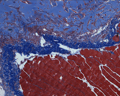

This is using a tri-chrome stain. The red is muscle and vascular tissue. The blue is adipose tissue and a special material we're using in our assay. The final color is black which stains the nuclei, but this magnification isn't high enough to see them.

Comments

kenju said…
I used to see stuff like that everyday, when I was a cytotechnologist (back in the dark ages).
Shephard said…
What a stunning photo.
~S
rosemary said…
I think I would stain all blue....
srp said…
Fibrous tissue also stains blue in the tri-chrome stain. Hard to say for sure on this power, but that looks like skeletal muscle...not smooth muscle for sure, but would have to see the fibers a bit higher to completely rule out cardiac... a sight for these sore eyes indeed.

My personal favorite stain is the GMS.
srp said…
I was judging the siz of the muscle fibers by the size of the vessels.... Most smooth muscle would be smaller and in layers of the GI tract and other internal organs. This just had the "block squared off" look of skeletal... but I guess the magnification and the site of dissection would be the telling factor. Muscle all stains red with the tri-chrome anyway.. it is still PRETTY!

Popular posts from this blog

Nitroflex at home

flea!

ankles: the sequel